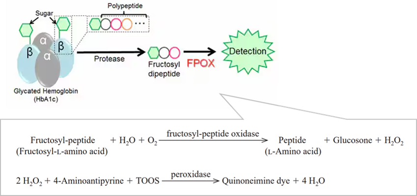
臨床検査において、フルクトシル-L-アミノ酸およびフルクトシルペプチドの測定に利用されます。また、プロテアーゼと併用することでHbA1cの測定に利用されます。(HbA1cは糖尿病の診断に検査指標として利用されています。プロテアーゼによって HbA1c から切り出されたフルクトシルペプチドまたはフルクトシル-L-アミノ酸をFPOXで測定することによって、HbA1cを定量化することが可能です。)
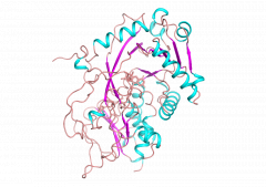
Img. Ref.:NAR 46: W296–W303 (2018)
| 由来 | recombinant E. coli |
|---|---|
| 系統名 | Fructosyl-peptide : oxygen oxidoreductase |
| EC 番号 | 1.5.3 |
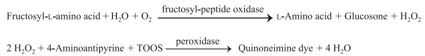
| 反応式 | Fructosyl-L-amino acid + H2O + O2 →→→ Peptide + Glucosone + H2O2 |